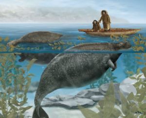
11
Nov 09

Deliciousness in Lab 3

Yum. Them sea urchins be REAL good eatin’ Uni or sea urchin is delicious gonad treat. There is nothing like some smooth gonads to make the day go by. However, not everyone likes these taste eggs or roe. Especially when it has been sitting in a tank for three weeks. We used Magnesium Chloride to kill our urchin unsuccessfully, so eating those oh, so edible ‘nads was out of the question. I decided that Thursday was not a good day to die. Well, actually it took tons of clove oil to kill our urchin. I think the clove oil would make uni taste like Christmas. In the pictures you can see that our uni was not so delicious looking.

Sea Urchin being euthanized by clove oil

Sea Urchin being euthanized by clove oil

Get your 'nads

Get your 'nads

dirty, sad uni in situ

dirty, sad uni in situ

This is what Uni should look like

This is what Uni should look like

Uni has increased in popularity and, therefore, as become a major industry. Harvesting of urchins in San Diego is harming the health of kelp forests. Interestingly, the kelp harvesting has been hurting the health of urchin population. These two industries  fight back and both causing man-supported urchin barrens and kelp forests.

Moral of the story? You should all go out and get yourself some uni. It’s delicious.

==============================================

Rate this review (click here)

==============================================

  • Share/Save/Bookmark

11
Nov 09

Migratory Bivalve?? No way man.

When people think of a bivalve organism, they should typically imagine a sessile animal with it’s shell partially or fully buried in the sand and feeding through its elongated siphon which filters out plankton in the water column.

A scallop, the only migratory bivalve organism, seems to defy many stereotypical morphological features that are endemic to the class Bivalvia.

Obviously since they are not sessile, they have had no need to develop a way to anchor themselves in one spot. Infaunal bivalves such as clams, have developed a foot that elongates and helps them bury themselves in soft substratum. Similarly, epifaunal bivalves such as mussels have developed byssal threads that hold them in place. Because scallops have no need for these mechanisms, its lifestyle differs from your typical bivalve.

After watching everyone torture the poor Spiny Pink Scallops (Chlamys rubida) in the sea tables in lab 3 yesterday, I became fascinated by the way they are able to swim.

I did find a couple interesting papers about scallop swimming dynamics.

http://www.sciencedirect.com/science?_ob=MImg&_imagekey=B6T8F-3W2516W-H-3&_cdi=5085&_user=582538&_orig=search&_coverDate=10%2F15%2F1996&_sk=997969997&view=c&wchp=dGLbVzW-zSkWz&md5=7db278f9ea08417da9054d64ba1df863&ie=/sdarticle.pdf

This first one is an interesting study on scallop body size and how it effects swimming trajectory in different flow speeds. They found that in higher flows smaller scallops always had a backwards net swimming trajectory in both their active swimming period and passive sinking period. Larger scallops made more progress in their active swimming period but were still at the mercy of the current in their passive sinking period. These results are fairly predictable but it’s cool to see swimming dynamics rigorously quantified.

http://journals.cambridge.org/download.php?file=%2FMBI%2FMBI83_04%2FS0025315403007847a.pdf&code=b40daa7be81a9e48bb451e4f5abcf862

This is a study on the effects of barnacle encrustation on scallop swimming dynamics. They found that barnacle encrustation negatively effects active swimming efficiency in both height and distance. Also they found that scallops encrusted with barnacles required more energy to swim a given distance. In the energetics studies they did, they found that there was no detectable difference in aerobic energy, but there was a difference in anaerobic energy required. Basically, they respired the same amount of oxygen, but more anaerobic nutrients were required by the barnacle encrusted scallops.

Another interesting (and relevant to our class!) thing I found in this paper is that while barnacle encrustation decreases the drag coefficient (Cd), sponge encrustation does not change it very much. So when our Chlamys rubida were frantically evading the Pychnopodia, the encrusting sponge should not have been making a very bid difference in drag.

Hoorah! Go Scallops!

-S.L.

==============================================

Rate Sean's blog (click here)

==============================================

  • Share/Save/Bookmark

11
Nov 09

Tête-á-tubefoot

“Those things don’t move, do they?”  This is one of the most common questions I hear at the Seattle Aquarium from guests crowded around the intertidal touch tanks.  The object to their subject is invariably the humble echinoderm.  ”Yes,” I inform them, “they certainly do move.”  ” But HOW?”  Ah, now we have hit upon the heart of the matter, or rather, the foot.  How DO they move?  Surely the seastar, sea urchin and cucumber are simply subject to the push and pull of the tides, going where the water flows without a care for how they get there.  Well, anyone who has ever tried to pull a Pisaster off the rocks will tell you otherwise.  They know that these creatures are not only capable of moving, but many are capable of NOT moving, clinging to surfaces with a kung-fu grip. So what is the secret to their pedal prowess?  Well, the mechanism is one which man has employed in his endeavors since ancient times.  The answer simply put, is hydraulics.

So far as we know, man invented the hydraulic pump sometime around the 3rd century BCE.  And the echinoderm?  Well, they have been employing hydraulic power since the Cambrian period, roughly 525 million years ago.  Some might say they beat us to the technological punch.  So how exactly does this unlikely water vascular system (as it’s called) actually work?  The answer will vary by species but there are relative similarities between all members of Echinodermata which are ever present…and amazing.

Cucumber Tube Feet

Cucumber Tube Feet

The basic structure of this incredible system consists of a few ’simple’  parts.  Firstly, a madreporite (also called hydropore), which acts as a basic incurrent/excurrent and pressure regulatory portion for the water vascular system.  It functions to equalize pressure of the system with the ambient water pressure, much in the same way SCUBA equipment adjusts air pressure to match depth.  In some echinoderms (seastars & urchins) this madreporite is located on the exterior of the animal, but in the sea cucumber, this pore is internal and is able to regulate pressure via the compression of soft tissues.  Next we come to the stone canal, a short tube which leads from the madreporite to the ring canal, the hub of water distribution for the rest of the body.  Attached to this ring canal are a number of radial canal, usually numbering five, reflecting an echinoderms pentaradial symmetry.  Also attached to the ring canal you will often find polian vesicles.  These vesicles are blind sacs which hold excess water under pressure and may be utilized to add more fluid to the system if it is ever needed.  Finally, we get to the crux of the matter, the all-important tube feet!  The tube feet are fleshy stalks which may or may not have suction cup-like ends and are used to grasp, pull, hold, move and otherwise manipulate the environment and their position within that environment.  Muscular and/or elastic canals act to operate these structures, and some may be associated with bulbs and ampullae that are capable of activating tube feet by local water pressure.  An ingenious system for locomotion if ever there was one.  Amazing!

Lucky for me, most guests at the Aquarium prefer demonstrations to wordy descriptions.  So in answer to their question I will simply find the nearest seastar, and gently flip it on its ‘back’.  ”Watch.”

==============================================

Rate Ross's Entry

==============================================

  • Share/Save/Bookmark

10
Nov 09

In the name of science..

What does this phrase mean? In some cases I think it serves as a blanket excuse for actions that would not otherwise be condoned, and in others, a legitimate cover for things that NEED to get done.  Who decides under which of these two categories “science” falls?  Sean was telling me last week about a class he took in the ethical/quick/most painless method for killing lab animals.  On one level I’m glad that they’re teaching this kind of stuff to the future scientists of the world, but on the other hand, I’m perturbed that so much of science includes using lab animals that there is a course in it.  In the case of the dissections we did on Thursday, I personally didn’t find the benefits of the dissection worth the deaths of the animals we dissected.  Sure, it was kinda cool to examine the digestive tracts of a crab under a microscope, but my background in marine biology is so limited that for the most part I had no idea what I was looking at (the guide we were provided was minimally helpful to me, no knock on it but I think it was written for people that had a basic familiarity with crabs).  Nor will I be applying this knowledge of a crabs insides to anything that I will be doing in the future.  What’s done is done, but I wouldn’t do it over again if the opportunity came up.

One thing I distinctly remember from this lab is catching the crab and preparing it for dissection.  We chose to use a net to catch him because he fought like hell to avoid capture, snapping his pincers and scuttling every which way.  It made me really sad to see that we would be dissecting something so full of life and vigor.  And then when we couldn’t anesthetize him properly, we decided to kill him by breaking him in half over the edge of the table.  I think its a little bit messed up that don’t have rules regulating the killing of invertebrates.  For my independent research project I’m doing work on some of the small farms on the island and one of them, Sweet Earth, is slaughtering turkeys and chickens in the coming weeks.  I’ve been invited to help out and I think I will if only to better understand something I’ve taken for granted up to now.  A greater appreciation for death might be the only thing I remember about this lab in 5 years, so I guess in that sense it was worth it.

tay

==============================================

Rate this review (click here)

==============================================

  • Share/Save/Bookmark

09
Nov 09

Mollusc Art – Phil

Now that Jessica has stolen my thunder by doing a great job of describing the crystalline sac and crystalline style, that fabulous organ in some mollusks that spins around in the sac, I none-the-less have to acknowledge her ability as an artist.  How she took an ordinary mussel that looked like this clam disection and turned it into this work of artclam disection-4is certainly beyond this non-artist.  Meanwhile, Brit and I were busy dissecting a clam and looking at pieces under the dissecting microscope.  And for those of you who have yet to read Jessica’s blog, the crystalline sac cut open with style inside looked like this on our specimen.clam disection-2 In the first photo above it’s the orange structure in the upper left.

Now on to a new topic (as long as Emily opened it up to non-lab entries.)

It was mentioned in class how some nudibranchs feed on Cnidarians and use their nematocysts.  Turns out that this may be important for biotechnological research.  The Cnidarian venoms are of interest because an estimated 150 million people are exposed to jellyfish stings yearly.  Preparations from nematocysts maybe useful in developing repellents to stings.  The question is how to harvest these nematocysts.

Aeolid nudibranchs have evolved over millions of years resulting in a lean mean Cnidarian eating machine.  Once eaten, the undischarged  nematocysts are divided and sent in one of two directions.  Some nematocysts go to the tips of the cerata by a process called foreign organellar retention where they serve a defensive mechanism.  The majority of the nematocysts are discharged in the nudibranchs’ feces.  Current research is developing processes to harvest the nematocysts from the nudibranch feces.  In the study, an average of 150,000 active nematocysts were excreted per day.  Using this process and using the nudibranch Spurilla neapolitana, it provided direct access to the entire range of venoms from four species of Cnidarians.  The authors concluded that their methods would help advance the research on the use of Cnidarian venoms as a source of marine natural products.

The above information was from the article Active Nematocyst Isolation Via Nudibranchs by Ami Schlesinger, et.al., Marine Biotechnology (2009),11:441-444.

spur_3

Can you imagine spending your day harvesting feces from the above Spurilla neapolitana and removing the 150,000nematocysts?  Welcome to biotech research.

Enjoy that thought,

Phil

==============================================

Rate this review (click here)

==============================================

  • Share/Save/Bookmark

08
Nov 09

Where have all the Gonads gone?

Question: What is worm-like in flexibility, orange and textured, and feeds off deposits on the bottom of the sea floor?

Echinodermata – Parastichopus californicus is exactly that… and more!

P1000492r

Parastichopus


In our dissection of this unfortunate sea cucumber Ross and I were able to explore the surprisingly simple inner workings of the sea cucumber. And by simple I mean the few parts that make this a living organism, not that the parts themselves are simple. What seemed like forever to sedate the poor creature in both mag-chloride and clove oil we finally just had to go for it and cut into it. We started at the mouth, which we learned may not have been the best approach, but how were we to know? Shortly after cutting into the cucumber the poor Parastichopus attempted its defense mechanism and eviscerated its guts for us to see. Most sea cucumbers can regenerate the internal organs that they expel, however we had other plans for this invert and proceeded to explore its internal workings.

P1000477r

Cutting it open

P1000479(1)r

evisceration of the guts


P1000484(1)r

Guts!


Our sample consisted of Buccal tentacles, Tentacular ampulla, a Calareous ring, polian vesicle, esophagus, intestine, hemal plexus, and respiratory tree, with podium covering the outside. The only thing we were missing was the Gonads. Where were they? Was the cucumber not of maturity, are they only seasonal I wondered? My knowledge of marine inverts has yet to be expanded until this point. As it turns out, Parastichopus eviscerates its gut contents between October and November every year and regenerates new ones. Spawning takes place in late summer, Parastichopus is a broadcast spawner. Sill, why no gonads in this specimen? I’ve found out through this process of gonad research that they have a closed circulatory system and hemoglobin, but no heart, water internal vascular system, and a calcite internal skeleton. They also have a 5 banded mussel wall structure that makes them part of the Echinodermata phylum like sea-stars and urchins, this is called pentaradial symmetry. They can reach up to 50 cm but typically are only around 24 cm -40 cm, are gonochoristic, and they can live up to 8 years if they are lucky enough to escape commercial harvesting. Sushi anyone? I also saw a recipe for sea cucumber chowder… yum? Apparently there is a huge market for sea cumber. I still haven’t found out about those gonads… Ah ha! The California department of fish and game has some information for me. They say that when the Parastichopus undergoes its seasonal visceral atrophy the gonads, circulatory system and respiratory tree are reabsorbed and shrink during the process of gut degeneration. They say that juveniles also undergo this process, but have not yet developed gonads. Could this be a victory? I have now 2 hypotheses as to why we didn’t see gonads in our Parastichopus, 1) it was a juvenile and hadn’t developed gonads yet (this one seemed adult due to its size though) and 2) it was undergoing seasonal visceral atrophy and the gonads were not visible to us, (yet other organs were visible).

P1000491r

Another species of sea cucumber


An interesting little factoid that I stumbled upon while doing research on the gonads was that sea cucumbers have dial and seasonal cycles of movement and metabolic activity. This made me think of the Nucella experiment and their tidal cycle. Correlation? We’ll have to find out!

 Thats it for now!

Liza

==============================================

Rate this review (click here)

==============================================

  • Share/Save/Bookmark

21
Oct 09

Getting hot in here

Last Tuesday we went to Cattle Point to catch the low tide at 6 in the morning.  We were all a little bit groggy from the early hour but the morning chill and the variety of the organisms inhabiting the intertidal zone were more than sufficient to spring us into alertness.  Emily turned over a rock to reveal little critters scrambling to get away from the light of the flashlights (the sun had yet to rise so we were using lights).  We also discovered some mussels grouped in tight circles and, of course, the omnipresent barnacle.  The mussels were being monitored by a temperature gauge that was affixed to the rock by what looked like silly putty, but in reality acted as a layer of insulation for the button-like instruments.  They looked like the tiny circular batteries for watches and exuded the same silvery metallic sheen.  Moose explained to some of us the rationale behind gathering data from spots like this year after year after year.  Apparently mussels are subject to high environmental stress from the changing temperatures and endure varying conditions that would kill most other organisms.  Interesting fact: the mussels of the Northwest are actually subject to higher heat than the mussels of Southern California.  This is because the high tides in Socal come during mid-day, keeping the mussels wet and cool during the hottest part of the day.  The Northwest is the opposite, and the low tides during mid-day expose the mussels to high heat and drying factors during the day.  Apparently the data collected over the years is matched with mortality rates among mussels and analyzed.  Mussels are amazingly resilient creatures.  A short 3 hours outside and I was shivering in what was probably somewhere around 40 degree weather.  They routinely survive exposure to temperatures much lower.  My Northface jacket just wasn’t cutting it.  Nature has got human technology beat.  Its like the site that Ross was showing us today on bio-mimicry.  Human hubris likes to pretend that we’ve moved past nature, conquered or tamed it, but we’re still infants in the biological timescale.

==============================================

Rate this review (click here)

==============================================

  • Share/Save/Bookmark

19
Oct 09

Cattle Point & Macro vs Micro Zonation Study Utility

CattlePoint_01(brit)

Cattle Point’s obvious biogeographic zones as well as the many species-level interactions framing these organic equilibrium states combine to highlight (for me anyway) a significant focal tension in biological and environmental sciences.

At the macro scale, astute longterm observations of rocky North Pacific shores loan themselves to teaching lessons that easily divide the intertidal into 3-4 zones: supralittoral fringe, upper midlittoral, lower midlittoral and infralittoral fringe. And, in places like Cattle Point, we define these macro-level divisions by pointing to the interactions of the Fucus, the Balanus glandula, the Mytilus, the Nucella lamellosa to showcase their existence. At the same time, those of us interested in the specific behaviors and biological processes of a small sampling of some particular species often choose to anchor our questions and answers in stress-level gradient assumptions about these same macro-level littoral zones.

A referential mobius strip.

Good scientific investigation—or, maybe better to say good communication and application of scientific investigation—requires a conceptual fluidity that moves both you and your audience back and forth along this infinite interchange. For example, the egg ribbons of the Archidoris montereyensis nudibrachs found at Cattle Point (yellow blobs pictured above) have been studied by Biermann et al. (1992) here at FHL to assess (among other things) whether UV radiation impacts on embryo survivorship result in limits to species distribution. This line of inquiry is an invitation to extend the discussion and conclusions of prior inquiry regarding UV impacts on planktonic larvae to benthic organisms.  And (although probably a very long stretch for this particular study) could maybe even point us down a path to describing and predicting potential ecosystem impacts caused by increased greenhouse gas emissions.

That’s useful right?

Its such a precarious tightrope in my mind… between the rigorous study of something that could potentially be viewed as “minutia” and the line of dots leading to a concept of “maximized utility”.   It’s just so easy to end up either stretching that tightrope line too far or not far enough.

This paper is a good case study for what I’m talking about (and also helps bring this post back to the zonation theme of the field trip!)

What do you guys all think of the monitoring method proposed and the rationale behind it?

==============================================

Rate Brit's review (click here)

==============================================

  • Share/Save/Bookmark

19
Oct 09

Look, ma–it’s almost terrestrial!

Here’s a lesson in not procrastinating: by this point, everybody else has already quite effectively covered the various awesome species of algae that Emily enlightened us to at Cattle Point.  But, luckily for me, the species I found most interesting was not an alga.  Unsurprisingly, it was Phyllospadix scouleri, surfgrass (see Sucia blog re: one-trick pony).

Surfgrass seems to be a pretty unique plant, both as terrestrial and marine plants go.  It’s one of the only angiosperms that can withstand being battered by the harsh conditions of the intertidal (hence the common name “surfgrass”), and also has a unique reproduction system.  A dioecious plant, surfgrass produces barbed seeds (below) which entangle themselves into nearby red algae and germinate immediately.  This is probably the coolest biology factoid I have heard.  Or at least the coolest one that comes to mind.

Barbed seeds (not fully developed) and sheath.

Barbed seeds (not fully developed) and sheath.

Surfgrass (far right) next to red algae next to barnacles.  Also a good example of zonation.

Surfgrass (far right) next to red algae next to barnacles. Also a good example of zonation.

But, even more interesting than that (“Not possible!” I hear you cry) are the results of a 1995 study by Susan Williams examining the distribution and ratios of sexes in Phyllospadix scouleri.  Williams was interested in the previously noted strong female bias among populations of Phyllospadix, as, though males are scarce, pollen is not.  There is a theory in ecology that in dioecious plants, the more energy-intensive sex to produce will be less competitive, thereby skewing the sex ratios.  Williams found that male reproductive organs were only slightly, but not statistically, higher than female reproductive organs, thereby demonstrating how even slight energy allocation differences can result in dramatic effects at the population level.  There is also a marked difference in the distribution of sexes; within the surfgrass bed, females are found in shallower waters while males prefer the deep end of the pool.  There are a few hypotheses as to why males are more competitive at deeper depths, including that the more continuous submersal facilitates pollen dispersal, and that, for unexplained reasons, the males are less strongly attached than females and therefore do better in less turbid subtidal zone.   Females, on the other hand, are fitter under more intense light conditions.

-Sarah

PS Just for kicks…

IMGP0009

==============================================

Rate this review

==============================================

  • Share/Save/Bookmark

19
Oct 09

Dude, What’s Up With That Sea Star?

Last week we went out to the rocky intertidal, specifically Cattle Point.

I always like seeing patterns of zonation, watching the lichens descend into algae and anemones, plunge into deep water and mussels and kelps, and seeing a more exposed coast and who likes to live there versus a sheltered coast (sometimes within a two minute walk from each other). If you like finding patterns, the rocky intertidal is a good place to be.

That said, what caught my attention this time was a little red sea star we found in a pool filled with the surf grass Phyllospadix scouleri.

Normally I’d look at it and say, “Oh. Yep, that’s Henricia,” and move on. But just as the sun was starting to really get up in the sky (and all of us were really beginning to get hungry) I could see how the surface of the star appeared like it was bubbled up with small glass beads.

When neither Moose nor Emily could tell us why, Ryan pulled a jar out of his trench coat and we packed it on home to look at.

My possible theories included:

  1. Parasite! Let’s be honest, I was hoping for a parasite because I think they’re just about my favorite thing in biology after learning so much about them this past summer.
  2. Disease or some kind of malformation–seemed possible.
  3. Predation: something had eaten off of his surface. This seemed possible because I know Harlequin shrimp in the wild will often keep a sea star at their mercy for weeks, eating one ray at a time, keeping them flipped over, feeding them food. By the time the shrimp is back to the ray it started on, there’s a good change it’s already regenerated a good deal of it (depending on how much food the shrimp gives it). It wouldn’t have surprised me if something was just picking off the delicious tube feet from sea stars.

Since looking at some others we had cruising around in the sea table in the lab, I’ve come to the conclusion that it’s much more likely this is on all of them–the specimen we found at Cattle Point was particularly large, so I think that’s why it seemed so unusual. They don’t have any pedicellariae according to literature I’ve seen so far, which seems to coincide with the occurrence of these strange tube-like holes on their surface.

I also have to face the possibility that maybe they always look like that and I haven’t taken the time to be a good observer and check for these baubles on each of the ones I’ve seen–I’ve written them off as soon as I saw them as “just another Henricia.” This is a little upsetting because I try and pride myself on taking the time to really look at an organism, rather than seeing what I think must be there and moving on before I’ve had time to go beyond my preconceived notions.

So, lesson learned. Even if I’ve seen it before, I should take the time to get a better understanding of how it looks, works, and functions in its environment. If anyone knows why, exactly, their holes end up looking bubbled and solid (still clear, however) I’m really interested in continuing to figure this out.

In the meantime, time to study more for this Sociology quiz.

–Meghan

==============================================

Rate this review (click here)

==============================================

  • Share/Save/Bookmark